Analyze Your Organoid
Upload a brightfield microscopy image to extract morphology features and run quality control checks.
Upload Image
Brightfield microscopy images of neural organoids (JPEG, PNG, or TIFF)
Drop your organoid image here
or click to browse
Try a Sample
Don't have an image? Analyze one of our dataset samples from each cell line.

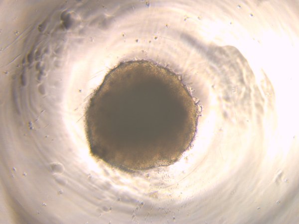
Wildtype (wt2D)
Healthy control, Day 10
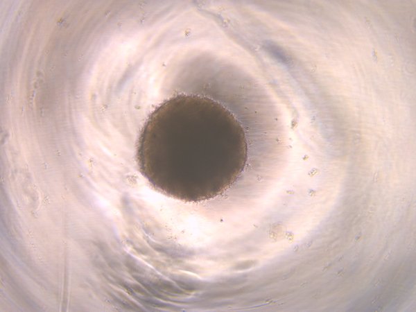
Disease Model A1A
TUBA1A mutation, Day 10
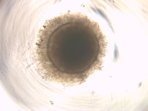
Disease Model B2A
TUBB2A mutation, Day 10
Disease Model TH2
TH mutation, Day 10
How It Works
Your image is processed through our morphology pipeline: adaptive thresholding for segmentation, connected-component analysis for object isolation, scikit-image for morphological feature extraction, and gradient-based texture analysis. Quality control evaluates focus sharpness (Laplacian variance) and signal-to-noise ratio. All processing is done server-side; no images are stored after analysis.